For the person anticipating scoliosis surgery, it is confusing
and sometimes troubling to learn of the wide variety of instrumentation
systems that are in use today. Why, the patient wonders, are there
so many? How are they different? Which one is best? That last question
is the easiest to answer. The fact is there is no one "best"
instrumentation for every patient or for every physician. In planning
the surgery, the physician takes a number of factors into account:
the location and magnitude of the curve, the degree of rotation,
the extent of deformity of the individual vertebrae, the rigidity
or flexibility of the spine, the density of the bone, and the size
of the patient. In addition, the physician may have a personal preference
and skill for working with one instrumentation or another.
The Purpose of Spinal Instrumentation
The purpose of the spinal instrumentation is twofold: First, it
enables the surgeon to reduce, adjust etc. the curvature to some
degree. To keep the curve from progressing, the surgeon performs
a spinal fusion and may utilize bone graft from the hip, bone bank,
collagraft bone substitutes, etc. Eventually, the grafted bone fuses
into a solid bone mass, and the vertebrae are permanently immobilized.
However, this takes time-up to a year or more for adults. During
this period, the instrumentation fulfills its second purpose: the
metal rods make the spine stiff and hold it still so that the fusion
can set. Once the fusion is solid, the instrumentation has done
its job and could be removed, although it is usually left in place.
The instrumentation will eventually fatigue and fail if a solid
fusion is not achieved
What follows is an explanation of some of the more popular systems
in use today:
Harrington Rod
Harrington-Scoliosis surgery was revolutionized in the early 1960's
with the introduction of the Harrington
Rod, designed by Dr. Paul Harrington, It was the first device
designed to straighten and immobilize the spine from inside the
body. It was so successful that it remained the "gold standard"
for scoliosis surgery for over 20 years.
The Harrington system achieves correction of the curve by stretching
or distracting the spine. The straight rod, containing a ratcheting
mechanism, is positioned along the inside or concavity of the curve.
It is attached to the spine with two hooks: one set into the vertebra
at the top of the curve, the other into the vertebra at the bottom
of the curve. Then, employing the ratcheting mechanism, the surgeon
stretches the spine to straighten the curve. Since the rod is attached
in only two places, it is necessary for the patient to wear a brace
after surgery to achieve more secure immobilization of the spine.
Even so, the vertebrae between the hooks sometimes fails to fuse
solidly. The most important drawback to the Harrington, however,
is that it allows little restoration of the normal contours of the
spine when viewed from the side. This includes the normal outward
curve at the top of the spine (kyphosis) and the normal inward curve
at the bottom (lordosis), which are typically distorted by scoliosis.
Luque-To achieves a more stable,
stronger fixation, Dr. Eduardo Luque of Mexico City devised the
Luque implant in the early 1970's. Two flexible L-shaped rods are
placed on either side of the spine. The rods are contoured or bent
to conform to the curve, and wires are threaded through the spinal
canal at each vertebral level. The wires are then twisted around
the rods on either side of the spine. The rods apply pressure on
the spine to correct the curve. Because there are multiple points
of fixation with the Luque technique, the patient generally does
not have to wear a brace after surgery as with the Harrington Rod.
However, since the wires pass through the spinal canal, this system
poses a greater risk of neurological damage than other systems.
Luque rods or variations on the Luque technique are still often
the preferred instrumentation for neuromuscular curves.
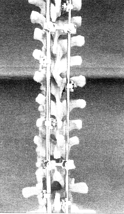
Multiple Hook and Contourable Rod Systems
In 1984, a new concept in spinal instrumentation developed by Drs.
Yves Cotrel and Jean Dubousset in France, was introduced in this
country. The Cotrel-Dubousset (CD)
instrumentation involves the use of two flexible rods and multiple
hooks as do both the TSRH developed
by the Orthopaedic staff at The Texas Scottish Rite Hospital, and
the Isola technique, designed by
Dr. Marc Asher and Dr. Charles Heinig and engineers Walter Strippgen
and Dr. William Carson. All of these multiple hook and contourable
rod systems deal with a problem of scoliosis which was not addressed
by earlier systems. When the spine curves sideways, the vertebrae
rotate towards the concavity of the curve. Since the ribs are attached
to the spine, they are dragged along and splayed out on the convex
side of the curve and compressed on the concave side. (This is what
creates the rib hump seen in a thoracic curve.)
With all of these systems, the surgeon bends or contours the rods
to conform to the desired profile. The rods are positioned on either
side of the spine and affixed to the vertebrae with multiple hooks
and sometimes screws as well. The rods themselves are joined to
each other by transverse rods or connecting devices.
The most important facet of the C-D, TRSH, or Isola is the ability
to control not just compression or distraction and not just scoliosis
correction but also to build in correction of lordosis or kyphosis.
The multiple hook and contourable rod systems differ from each other
mainly in the way the hooks are attached to the rods. The C-D, for
example, uses a set screw; the TSRH, a nut and bolt arrangement,
and the Isola, a drop set in screw. Basically, however, they work
on the same principle and accomplish the same ends. Because they
provide a very stable fixation, they usually do not require the
wearing of a brace.
Systems Used in Frontal Procedures
Sometimes if the curve is rigid or very severe, the surgeon will
perform an anterior procedure (going in from the front of the body)
in which he first removes some of the discs-the rubbery shock absorbers
located between the vertebrae. This makes the curve more flexible
and easier to correct.
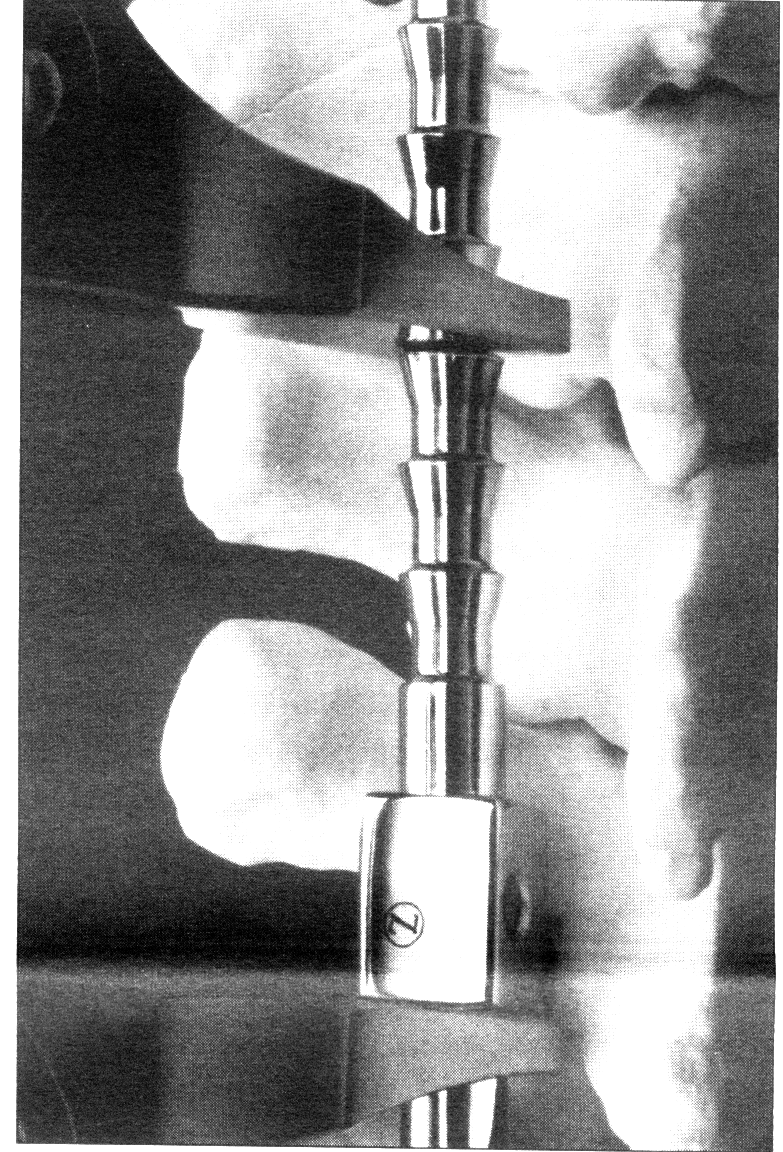
One system specifically designed to be used anteriorly is the Zielke
instrumentation, developed by Dr. Klaus Zielke of Germany. It
uses a flexible rod attached to the convexity or outside of the
curve with screws. Correction is achieved by compression of the
curve. Other systems which can be used for an anterior procedure
include the Dwyer, which is similar to the Zielke, the TSRH and
the Isola, as well as others. An anterior fusion may be performed
alone or frequently in conjunction with a posterior fusion.
There is significant ongoing research and development for spinal
fixation systems resulting in many new product introductions. The
spine instrumentation devices presented in this article are representative
of the multitude of products currently available. The NSF does not
recommend or promote any particular device and strongly suggests
that patients address any questions they may have about instrumentation
with their physicians.
"...there is no one "best" instrumentation for
every patient or every physician."
Back to Medical Updates